
GM1 Gangliosidosis Type 2b – Juvenile
- Type 2b symptoms typically begin between 2 and 5 years of age
- Juvenile GM1 accounts for approximately 26.1% of all GM1 gangliosidosis cases in the Cure GM1 Census.
- Primarily neurological: progressive dystonia, speech loss, and cognitive decline
- Does not cause the enlarged liver/spleen, cherry-red spot, or coarse facial features seen in Type 1
- Survival typically extends into the patient’s 20s or 30s
- No approved disease-modifying treatment exists, but the research pipeline is more active than ever
GM1 gangliosidosis (all subtypes combined) occurs in approximately 1 in 100,000 to 200,000 live births worldwide.
How Juvenile GM1 Differs from Other Forms of GM1
Researchers understand GM1 gangliosidosis as a spectrum, with subtype determined by residual enzyme activity. Below, we outline juvenile GM1 (Type 2b) and other subtype symptoms.
|
*Ave based on current literature |
GM1 Type 1 (Infantile) |
|
Onset |
Birth to 6 months* |
|
Residual Enzyme |
<1-5% |
|
Progression |
Rapid |
|
Survival |
Avg. 2-3 years* |
|
Multi-organ involvement |
Severe |
|
*Ave based on current literature |
GM1 Type 2a and Type 2b (Late-Infantile / Juvenile GM1) |
|
Onset |
7 months to 5 years* |
|
Residual Enzyme |
Approx. 5-15% |
|
Progression |
Moderate |
|
Survival |
10 years to early adulthood* |
|
Multi-organ involvement |
Mild-moderate |
|
*Ave based on current literature |
Type 3 (Adult / Chronic) |
|
Onset |
Adolescence to adulthood* |
|
Residual Enzyme |
>10-15% |
|
Progression |
Slow |
|
Survival |
Decades* |
|
Multi-organ involvement |
Primarily neurological |
Type 2b is distinguished from Type 2a primarily by its later onset and slower progression. Children with Type 2b typically develop normally for the first 2-5 years of life before neurological symptoms emerge. The hallmark presentation is progressive dystonia and gait disturbance, rather than the hypotonia and rapid regression seen in Type 2a. Hepatosplenomegaly, cherry-red spots, and coarse facial features are absent.
Unlike Type 2a, odontoid hypoplasia is generally not a feature of Type 2b, which further distinguishes the two juvenile subtypes clinically. The slower pace of disease in Type 2b means that many affected individuals survive into their twenties or beyond, though with significant disability.
Signs and Symptoms of GM1 Type 2b
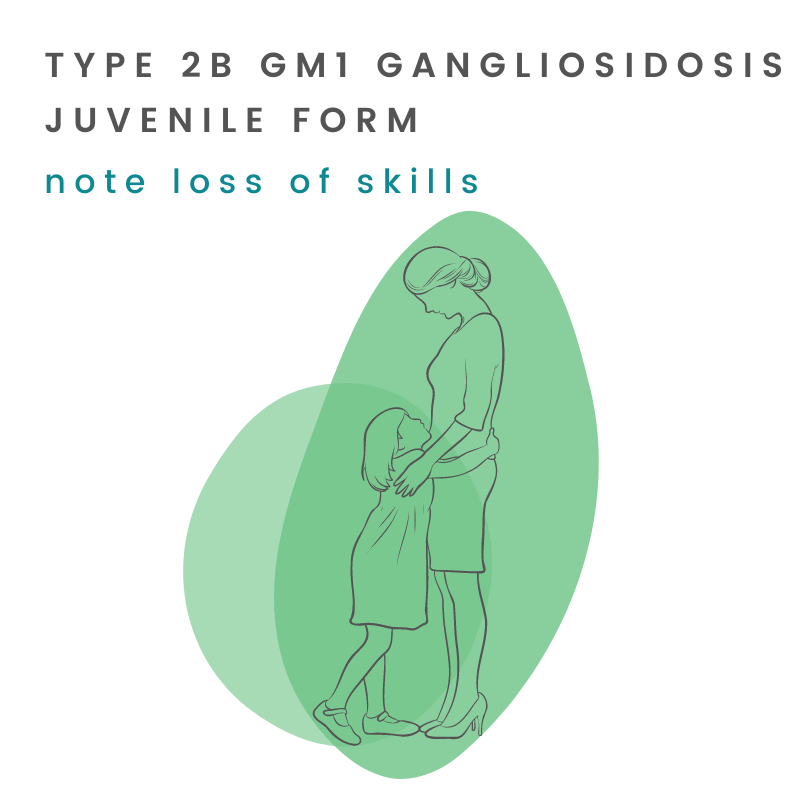
Symptoms of juvenile GM1 gangliosidosis typically emerge between 2 and 5 years of age, after a period of apparently normal early development. The disease progresses more slowly than GM1 Type 2a and primarily affects the brain and nervous system.
Early Neurological Signs (Ages 2-5)
- Gait disturbance: Unsteady or awkward walking is often the first sign noticed, reflecting cerebellar and extrapyramidal involvement.
- Dystonia: Involuntary muscle contractions causing repetitive or twisting movements. Dystonia is a hallmark of Type 2b and may begin in one limb before spreading.
- Speech difficulties (dysarthria): Slurred or increasingly difficult-to-understand speech, reflecting deteriorating motor control of the mouth and throat.
Progressive Features (Later Course)
- Intellectual regression: Gradual decline in cognitive abilities, including learning, memory, and attention, becomes apparent over time.
- Ataxia: Incoordination affecting gait, limb movements, and balance.
- Loss of independent ambulation: As dystonia and spasticity worsen, most affected individuals eventually lose the ability to walk independently.
- Seizures: Less frequent and later in onset than in Type 2a, but seizures do occur in a proportion of Type 2b patients.
- Spasticity: Progressive muscle stiffness develops over time, often in combination with dystonia.
- Feeding and swallowing difficulties: As bulbar function declines, dysphagia and aspiration risk increase; gastrostomy feeding may eventually be needed.
- Loss of language: Speech is typically lost over the course of the disease, though the timeline varies considerably among individuals.
What Is Notably Absent (Compared to Types 1 and 2a)
- No enlarged liver or spleen (hepatosplenomegaly)
- No cherry-red spot on eye examination
- No coarse facial features
- No odontoid hypoplasia (unlike GM1 Type 2a)
- Normal hearing in most patients
- No significant cardiac involvement in early disease
Average Juvenile GM1 Timeline
|
Age Range |
What Families Often Notice |
|
Birth to ~2 years |
Development typically appears normal. No obvious signs of disease in most children. |
|
2-5 Years |
Gait becomes unsteady or awkward. Dystonic movements may begin. Speech starts to become unclear. |
|
5-10 years |
Progressive dystonia and intellectual decline. Speech increasingly difficult. Some children develop seizures. |
|
10-15 years |
Loss of independent walking in many patients. Increasing dependence for daily activities. Swallowing difficulties may emerge |
|
15+ years |
Significant neurological disability; many individuals require full care. Respiratory complications become a concern. |
How is GM1 Type 2b diagnosed?
Doctors frequently misdiagnose juvenile GM1 gangliosidosis or diagnose it late. Because symptoms emerge gradually in previously developing children and lack the dramatic features of Type 1, they may mistake Type 2b for cerebral palsy, inherited dystonia syndromes, or other childhood neurodegenerative conditions. Diagnostic delays of several years are common.
The Diagnostic Pathway
- Enzyme activity assay: Measurement of beta-galactosidase activity in white blood cells (leukocytes) from a blood draw, or in skin fibroblasts from a small biopsy. Reduced activity (typically 10-15% of normal controls) confirms the diagnosis. This is the most important first-line test.
- Molecular (genetic) testing of the GLB1 gene: Identifies the specific mutations present and is essential for carrier testing, prenatal diagnosis, and distinguishing Type 2b from Types 2a and 3 at the molecular level.
- Brain MRI: Often shows progressive cerebral and cerebellar atrophy and white matter changes, though early MRIs may appear near-normal or show only subtle findings.
- MR spectroscopy (MRS): May reveal neuronal loss markers (reduced NAA) and glial activation markers (elevated myo-inositol), consistent with the natural history study findings.
Common Diagnostic Routes
- Pediatric neurologist evaluating progressive dystonia or unexplained gait deterioration in a school-age child
- Metabolic genetics referral after dystonia workup fails to identify a common cause
- Incidental identification through family history after a sibling is diagnosed
- Expanded newborn or childhood screening (if available)
- If you suspect GM1 in your child, an urgent referral to a metabolic genetics specialist or pediatric neurologist with lysosomal storage disorder experience is critical. An enzyme activity assay from a routine blood draw can confirm or exclude the diagnosis quickly.
GM1 Gangliosidosis Type 2b Prognosis and Life Expectancy
Juvenile GM1 gangliosidosis is a progressive and ultimately fatal disease, but its course is considerably slower than the infantile and late infantile forms. Many individuals with Type 2b survive into their 20s or 30s, though with increasing neurological disability over time.
The NIH 10-Year Prospective GM1 Type 2 Natural History Study (D’Souza et al., Genetics in Medicine, 2024) provides a detailed longitudinal data available on Type 2 disease progression. The study characterizes how brain atrophy, MRS biomarkers, and clinical function evolve over time in both GM1 Type 2a and GM1 Type 2b patients, and directly informs the design of upcoming treatment trials. See: PubMed
Survival is influenced by the pace of neurological progression, the frequency and severity of seizures, and the quality of respiratory and nutritional support. Respiratory failure, typically following aspiration pneumonia, remains the most common cause of death. There is real reason for hope: treatment approaches now in development, including enzyme replacement therapy, gene therapy, and substrate reduction therapy, may fundamentally change the prognosis for individuals diagnosed with Type 2b in the years ahead.
Learn how juvenile GM1 has impacted sisters Kinley and Kennedy who were diagnosed with GM1 after meeting typical developmental milestones until about 5 years old.
And, learn how Enzyme Replacement Therapy could help provide a bridge to promising treatment.
Management and Supportive Care Options for Juvenile GM1
No approved therapy currently stops or reverses progression of juvenile GM1 gangliosidosis. All management is supportive and symptom-focused.
Key Elements of Care
- Dystonia management: Medications such as trihexyphenidyl, baclofen, or tetrabenazine may reduce dystonic symptoms. Intrathecal baclofen or deep brain stimulation have been used in some patients with severe dystonia.
- Seizure management: Anti-epileptic medications are used as needed. Seizures in Type 2b are generally less refractory than in GM1 Type 2a, but management requires specialist oversight.
- Nutritional support: As swallowing coordination declines, gastrostomy feeding (G-tube) helps maintain adequate nutrition and reduces aspiration risk.
- Respiratory management: Chest physiotherapy and monitoring for respiratory infections. Respiratory support needs increase as the disease progresses.
- Physical, occupational, and speech therapy: Early and ongoing therapy helps maintain mobility, communication, and quality of life as long as possible.
- Neurology and genetics follow-up: Regular monitoring of disease progression and adjustment of medications and care plans.
- Educational and psychological support: Cognitive decline affects school performance and daily functioning; educational planning and psychological support for affected individuals and families are important components of care.
- Palliative care: Early integration of a palliative care team helps families navigate goals of care and symptom management across the disease course.
Research and Clinical Trials for Juvenile GM1
Type 2b is included in the active treatment pipeline that Cure GM1 Foundation is advancing.
Enzyme Replacement Therapy (ERT)
Cure GM1 Foundation is developing the first ICV (intracerebroventricular) enzyme replacement therapy for GM1, delivering recombinant beta-galactosidase directly to the cerebrospinal fluid to bypass the blood-brain barrier. This approach is modeled on the precedent set by Brineura (cerliponase alfa), FDA-approved for Batten disease via ICV infusion.
Gene Therapy
AAV-based gene therapy programs aim to deliver a functional copy of the GLB1 gene to brain cells. Results from Phase 1/2 AAV9-GLB1 gene therapy in Type 2 patients were published in 2025, with preliminary evidence of safety and biochemical signals. Type 2b patients, with their longer treatment window and greater remaining neurological reserve at diagnosis, may be well positioned to benefit from gene therapy approaches. AAV-based gene therapy update.
Substrate Reduction Therapy (SRT)
Azafaros’ nizubaglustat is entering a Phase 3 trial (NAVIGATE) targeting late-infantile and juvenile GM1/GM2 and NPC disease. Type 2b patients are included in this trial’s target population. Because Type 2b patients retain residual enzyme activity, reducing the rate of GM1 ganglioside synthesis through SRT may slow the pace of accumulation and neurological injury. Learn more about the NAVIGATE trial: ClinicalTrials.gov
Resources for Newly Diagnosed GM1 Gangliosidosis Type 2b Families
If your child was just diagnosed with juvenile GM1 gangliosidosis, you are not alone. This diagnosis is rare, and many families feel isolated and overwhelmed in the early weeks. Cure GM1 Foundation is here to help you navigate what comes next.
First Steps We Recommend
- Get to a metabolic genetics specialist. Academic medical centers with lysosomal storage disorder expertise are best positioned to manage this diagnosis. We can help you find the right center.
- Connect with other families. Parents and caregivers who have walked this path offer a kind of understanding and practical wisdom that no physician can provide. Cure GM1 connects families across the world.
- Ask about the natural history study and registry. Participation in research, even if no experimental treatment is available yet, helps scientists understand Type 2b better and brings effective treatments closer.
- Plan for educational and therapeutic support early. Cognitive and motor decline affect schooling and daily life. Early engagement with educational specialists and therapists can make a meaningful difference in quality of life.
List of Resources for GM1 Families
Join the GM1 Census
Contact Cure GM1
Frequently Asked Questions
GM1 gangliosidosis Type 2b, also called juvenile GM1, is a rare inherited neurological disease caused by reduced activity of the enzyme beta-galactosidase (due to two mutated copies of the GLB1 gene). It presents between 2 and 5 years of age, after a period of apparently normal development, with progressive dystonia, gait disturbance, and cognitive decline. Unlike Type 1 and Type 2a, it does not cause enlarged organs or cherry-red spots and progresses more slowly than the late infantile form. Survival typically extends into the patient’s 20s or 30s.
The key differences are age of onset, rate of progression, and specific neurological features. Type 2a (late infantile) presents between 7 and 24 months with rapid regression, hypotonia, and a mean survival of about 9 years. Type 2b (juvenile) presents between 2 and 5 years, after normal early development, with dystonia and gait disturbance as the dominant early features, and a much longer survival extending into the patient’s 20s or 30s. Type 2a also features odontoid hypoplasia, which is generally absent in Type 2b. The underlying biological difference is residual enzyme activity: Type 2b patients retain slightly more beta-galactosidase activity than Type 2a patients.
The most common early signs are gait disturbance (unsteady or awkward walking) and the beginning of dystonic movements, typically appearing between ages 2 and 5 after a period of normal development. Speech may become slurred. Intellectual regression, though present, is often subtle in the early years. Because these signs overlap with other childhood neurological conditions, diagnostic delays of several years are common.
No. Cherry-red spots are absent in Type 2b (and Type 2a). They are a feature of the most severe enzyme deficiency seen in Type 1 infantile GM1. The absence of a cherry-red spot in an older child with progressive dystonia does not exclude GM1 and should not discourage testing.
Diagnosis requires measurement of beta-galactosidase enzyme activity, typically from a blood draw (leukocytes). Reduced activity (approximately 10-15% of normal) confirms the diagnosis. Genetic testing of the GLB1 gene identifies the specific mutations and is important for family planning and distinguishing Type 2b from other subtypes. Brain MRI may show atrophy and white matter changes, though early scans can appear near-normal. Diagnosis typically follows referral to a metabolic genetics specialist or pediatric neurologist.
Survival typically extends into the 20s or 30s for individuals with Type 2b, making it substantially longer than Type 2a. However, prognosis varies considerably depending on the specific GLB1 mutations, the pace of neurological progression, and the quality of supportive care. Respiratory failure, typically following aspiration pneumonia, is the most common cause of death. Some individuals with Type 2b survive into their thirties.
Yes. Type 2b is included in the active treatment pipeline. Cure GM1 Foundation is advancing an enzyme replacement therapy program for GM1, with FDA regulatory engagement underway. Gene therapy studies (AAV9-GLB1) have published Phase 1/2 results in Type 2 patients. The NAVIGATE Phase 3 trial for nizubaglustat specifically targets late-infantile and juvenile GM1 disease, including Type 2b. We maintain an updated list of active research and trials: https://www.curegm1.org/gm1-clinical-trials-guide/
Yes. If a family has one affected child, siblings can be tested for GLB1 mutations or enzyme activity to determine whether they are also affected before symptoms begin. Pre-symptomatic identification is particularly valuable for Type 2b, because the wider treatment window means that future therapies initiated before neurological injury may have greater impact. Currently no country includes GM1 on its national newborn screening panel, but Cure GM1 Foundation is actively working to change this.
Connecting with the Cure GM1 patient registry also helps build the evidence base needed to evaluate future therapies.
Join the GM1 Census
Yes. Because GM1 is autosomal recessive, when both parents are confirmed carriers, each pregnancy carries a 25% (1-in-4) chance of being affected. Parents who have had one affected child are strongly encouraged to consult a genetic counselor to discuss carrier testing, recurrence risk, and options for future pregnancies including preimplantation genetic testing (PGT-M) with IVF, or prenatal testing.
Read Their Stories
Joey’s experience

“My daughter’s not able to tell me when she’s in pain, when she is hurting. I just have to guess. … I can just see her face. Just guessing what’s going on in her body.”
Ruth, mother of 20-year-old daughter living with Type 2b, juvenile GM1
Iris’ Experience

References
Brunetti-Pierri N, Scaglia F. GM1 gangliosidosis: review of clinical, molecular, and therapeutic aspects. Mol Genet Metab. 2008;94(4):391-396. https://www.sciencedirect.com/science/article/abs/pii/S1096719208001182
Regier DS, Kwon HJ, Johnston J, et al. MRI/MRS as a surrogate marker for clinical progression in GM1 gangliosidosis. Am J Med Genet A. 2016;170(3):634-644. https://onlinelibrary.wiley.com/doi/abs/10.1002/ajmg.a.37468
D’Souza P, Farmer C, Johnston JM, et al. GM1 gangliosidosis type II: results of a 10-year prospective study. Genet Med. 2024;26(7):101144. https://www.sciencedirect.com/science/article/pii/S1098360024000777
Kannebley JS, Silveira-Moriyama L, Bastos LO, Steiner CE. Clinical findings and natural history in ten unrelated families with juvenile and adult GM1 gangliosidosis. JIMD Reports. 2015;24:115-122. https://link.springer.com/chapter/10.1007/8904_2015_451
Kolstad J, et al. Natural history progression of MRI brain volumetrics in type II late-infantile and juvenile GM1 gangliosidosis patients. Mol Genet Metab. 2025;144(3):109025. https://www.sciencedirect.com/science/article/abs/pii/S1096719225000162
Nicoli ER, Annunziata I, d’Azzo A, Platt FM, Tifft CJ, Stepien KM. GM1 Gangliosidosis—A Mini-Review. Front Genet. 2021;12:734878. https://pubmed.ncbi.nlm.nih.gov/34539759/
Heron B, Batzios S, Mengel E, et al. A natural history study of pediatric patients with early onset of GM1 gangliosidosis, GM2 gangliosidoses, or Gaucher disease type 2 (RETRIEVE). Orphanet J Rare Dis. 2024;19:459. https://doi.org/10.1186/s13023-024-03409-1
MedLink Neurology. GM1 gangliosidosis. Accessed 2026. medlink.com
Published: May 2026
Cure GM1 does not prescribe medications or treatments. This information is being shared for educational purposes and discussion with your doctors.Visit us at curegm1.org
Help us find effective treatments and therapies so those living with GM1 can live long full lives
Donate to Cure GM1