
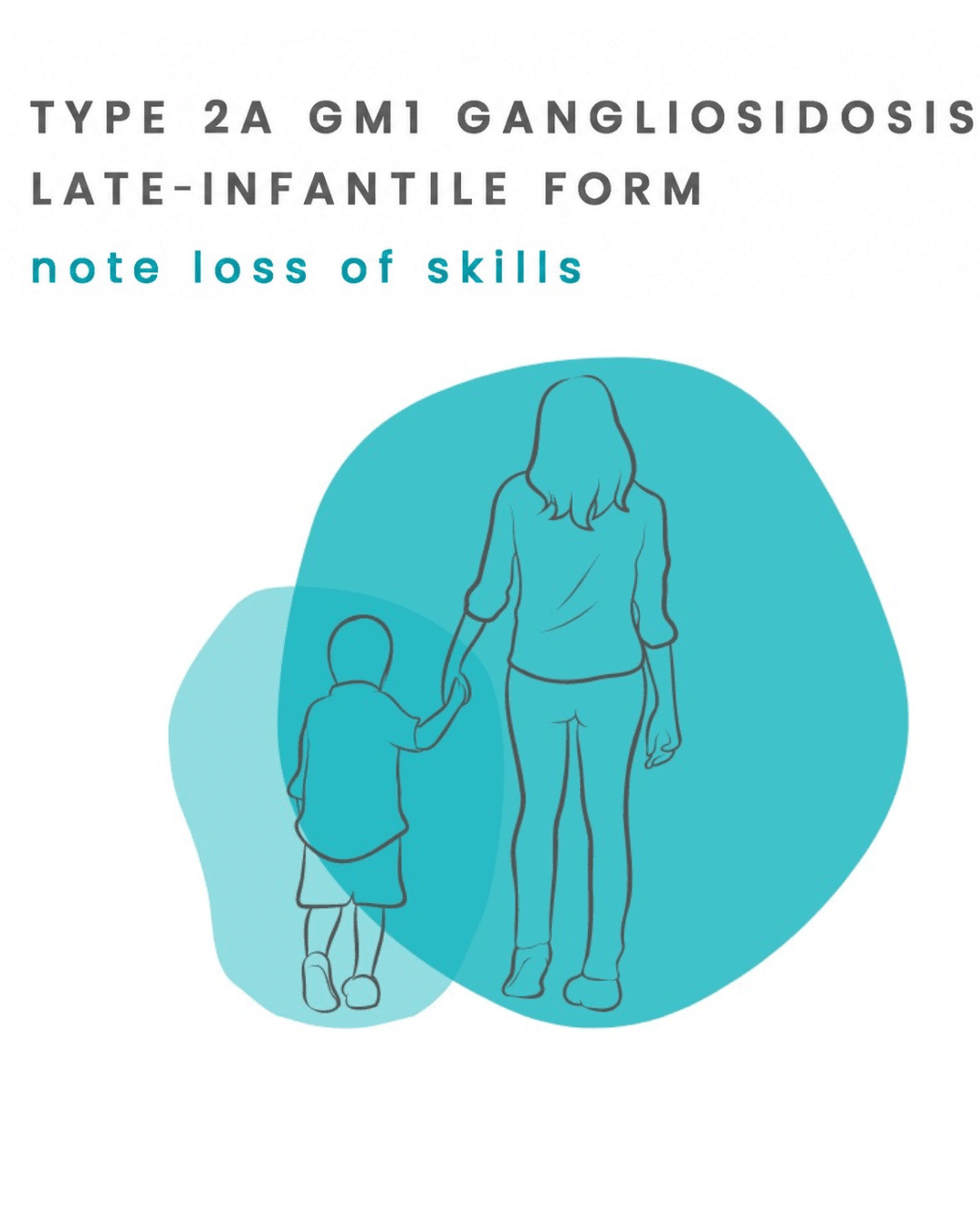
Late-Infantile GM1 Gangliosidosis (Type 2a)
- Type 2a symptoms typically begin between 7 and 24 months of age
- In the Cure GM1 Census, Type 2a registrants account for 18.8% of all GM1 gangliosidosis cases
- Primarily neurological: developmental regression, loss of language, ataxia, and seizures
- Does NOT usually cause the enlarged liver/spleen, cherry-red spot, or coarse facial features seen in Type 1
- Mean survival is approximately 9 years; some children survive into their early teens
- No approved disease-modifying treatment exists, but the research pipeline is more active than ever
What Is Late-Infantile GM1 Gangliosidosis (Type 2a)?
Late-infantile GM1 gangliosidosis, also called GM1 gangliosidosis Type 2a, is a rare, inherited, and progressive neurodegenerative disease. It is one of the two forms of “Type 2” GM1 gangliosidosis (the other being Type 2b, the juvenile form). It sits on the clinical spectrum between the rapid devastation of Type 1 infantile GM1 and the slower course of Type 2b.
Like all forms of GM1 gangliosidosis, Type 2a is caused by mutations in the GLB1 gene, which encodes the enzyme beta-galactosidase (beta-gal). When a child inherits two mutated copies of GLB1 (one from each parent), beta-galactosidase is severely reduced but not completely absent. This residual enzyme activity, typically around 5-15% of normal, is what distinguishes Type 2a from Type 1 (where activity falls below 1-5%) and largely explains the later onset and slower progression of Type 2a compared to the infantile form.
Without sufficient beta-galactosidase, a fatty molecule called GM1 ganglioside accumulates in neurons and other cells throughout the body, progressively impairing and destroying them. In Type 2a, this toxic buildup predominantly affects the brain and nervous system. The spleen, liver, and heart are largely spared, setting Type 2a apart from the multi-organ involvement that characterizes Type 1.
GM1 gangliosidosis is inherited in an autosomal recessive pattern: both parents must each carry one mutated copy of the GLB1 gene, and each pregnancy carries a 1-in-4 (25%) chance of an affected child.
GM1 gangliosidosis (all subtypes combined) occurs in approximately 1 in 100,000 to 200,000 live births worldwide. Type 2a (late-infantile) represents roughly 12-15% of all GM1 cases.
How Late-Infantile GM1 Type 2a Differs from Other GM1 Subtypes
|
*Ave based on current literature |
GM1 Type 1 (Infantile) |
|
Onset |
Birth to 6 months* |
|
Residual Enzyme |
<1-5% |
|
Progression |
Rapid |
|
Survival |
Avg. 2-3 years* |
|
Multi-organ involvement |
Severe |
|
*Ave based on current literature |
GM1 Type 2a and Type 2b (Late-Infantile / Juvenile) |
|
Onset |
7 months to 5 years* |
|
Residual Enzyme |
Approx. 5-15% |
|
Progression |
Moderate |
|
Survival |
10 years to early adulthood* |
|
Multi-organ involvement |
Mild-moderate |
|
*Ave based on current literature |
Type 3 (Adult / Chronic) |
|
Onset |
Adolescence to adulthood* |
|
Residual Enzyme |
>10-15% |
|
Progression |
Slow |
|
Survival |
Decades* |
|
Multi-organ involvement |
Primarily neurological |
How Is Type 2a GM1 Diagnosed?
Late-infantile GM1 gangliosidosis is frequently not recognized early. Because the disease lacks the dramatic multi-organ features of Type 1 (no enlarged liver, no cherry-red spot), it is more often mistaken for other causes of developmental regression, including cerebral palsy, autism spectrum disorder, or unspecified metabolic disorders. The median time from symptom onset to diagnosis in late-infantile GM1 has been reported at approximately 1.5 years.
The Diagnostic Pathway
- Enzyme activity assay: Measurement of beta-galactosidase activity in white blood cells (leukocytes) from a blood draw, or in skin fibroblasts from a small biopsy. Reduced activity (typically 5-15% of normal controls) is the confirmatory test.
- Molecular (genetic) testing of the GLB1 gene: Identifies the specific mutations present and is essential for carrier testing, prenatal diagnosis, and distinguishing Type 2a from Types 2b and 3 at the molecular level.
- Brain MRI: Often shows progressive cerebral atrophy, cerebellar atrophy (more pronounced than in Type 2b), and white matter changes. Early MRIs may be near-normal.
- MR spectroscopy (MRS): May reveal elevated myo-inositol (a marker of glial activation) and reduced N-acetylaspartate (NAA, a marker of neuronal health), findings that track with disease severity and have been validated as surrogate markers in a natural history study.
Common Diagnostic Routes
- Developmental pediatrician or pediatric neurologist evaluating developmental regression or failure to thrive
- Genetic counseling referral after concerning findings on clinical exam
- Expanded newborn screening (if available) or metabolic panel identifying abnormal results
If you suspect GM1 in your child, an urgent referral to a metabolic genetics specialist or pediatric neurologist with lysosomal storage disorder experience is critical. Time to diagnosis matters.
Average GM1 Type 2a Timeline
|
Age |
What Families Often Notice |
|
|
7-18 months |
|
|
|
18-24 months |
|
|
|
2-3 years |
|
|
|
3-5 years |
|
|
|
5 years plus |
|
Prognosis and Life Expectancy
Late-infantile GM1 gangliosidosis is a fatal disease, but its course is slower than the infantile form. A retrospective study of French patients (the largest published historical cohort, n=61, published in JIMD 2023) reported a mean overall survival of approximately 9.1 years (95% CI: 4.5-13.5 years) for Type 2a patients, substantially longer than the ~23-month mean survival for Type 1, but much shorter than the survival seen in juvenile (Type 2b) disease.
Survival is shaped by the pace of neurological progression, the severity of seizures, and the quality of respiratory and nutritional management. Respiratory failure, typically related to aspiration pneumonia, is the leading cause of death.
There is, however, real reason for hope. As in all GM1 subtypes, the research landscape is changing. Treatment approaches now in development, including gene therapy, enzyme replacement therapy, and substrate reduction therapy, may fundamentally alter what “prognosis” means for children diagnosed with Type 2a in the years ahead.
Current Management and Supportive Care
No approved therapy currently stops or reverses progression of late-infantile GM1 gangliosidosis. All management is supportive and symptom-focused. Full overview of where to find treatment for GM1.
Key Elements of Care
- Seizure management: Anti-epileptic medications are initiated when seizures begin. Seizure control in Type 2a can be challenging and may require multiple medication trials.
- Nutritional support: As swallowing coordination declines, a feeding gastrostomy (G-tube) is typically recommended to ensure adequate nutrition and reduce aspiration risk.
- Respiratory management: Chest physiotherapy, suction, and monitoring for respiratory infections. As the disease progresses, respiratory support becomes central to maintaining quality of life.
- Physical, occupational, and speech therapy: Early and ongoing therapy maintains function and quality of life as long as possible.
- Neurology and genetics follow-up: Regular monitoring of disease progression and management of emerging complications.
- Palliative care: Early involvement of a palliative care team helps families navigate goals of care and symptom management across the disease course.

Research and Clinical Trials: The Path Forward
Type 2a is included in the active treatment pipeline that Cure GM1 Foundation is advancing.
Enzyme Replacement Therapy (ERT)
Cure GM1 Foundation is developing the first ICV (intracerebroventricular) enzyme replacement therapy for GM1, delivering recombinant beta-galactosidase directly to the cerebrospinal fluid to bypass the blood-brain barrier. This approach is modeled on the precedent set by Brineura (cerliponase alfa), FDA-approved for Batten disease via ICV infusion.
Gene Therapy
AAV-based gene therapy programs aim to deliver a functional copy of the GLB1 gene to brain cells. Results from Phase 1/2 AAV9-GLB1 gene therapy in Type 2 patients were published in 2025, with preliminary evidence of safety and biochemical signals. Type 2a patients have a wider treatment window than Type 1 (because some neurological reserve remains at the time of diagnosis), which may make gene therapy particularly promising for this group. In-depth update on AAV-gene therapy for GM1.
Substrate Reduction Therapy (SRT)
Azafaros’ nizubaglustat is entering a Phase 3 trial (NAVIGATE) targeting juvenile GM1/GM2 and NPC disease. Because Type 2 patients retain some residual enzyme activity, SRT’s mechanism of reducing ganglioside synthesis may offer benefit by reducing the rate of accumulation.
Resources for Newly Diagnosed Families
If your child was just diagnosed with late-infantile GM1 gangliosidosis, we want you to know you are not alone. This diagnosis is rare, and many families feel isolated and overwhelmed in the early weeks. Cure GM1 Foundation is here to help you navigate what comes next.
First Steps We Recommend
- Join the GM1 Census. The GM1 Census helps families affected by GM1 gangliosidosis to share patient data that can guide research, improve diagnosis, and make our global community visible.
- Get to a metabolic genetics specialist. Academic medical centers with lysosomal storage disorder expertise are best positioned to manage this diagnosis. We can help you find the right center.
- Connect with other families. Parents and caregivers who have walked this path offer a kind of understanding and practical wisdom that no physician can provide. Cure GM1 connects families across the world. Check out our GM1 caregiver support group here.
- Ask about the natural history study and registry. Participation in research, even if no experimental treatment is available yet, helps scientists understand Type 2a better and brings effective treatments closer.
Helpful Links
Learn about Violet’s experience as a child diagnosed with late-infantile GM1.
Frequently Asked Questions
GM1 gangliosidosis Type 2a, also called late-infantile GM1, is a rare inherited neurological disease caused by severely reduced activity of the enzyme beta-galactosidase (due to two mutated copies of the GLB1 gene). It presents between 7 and 24 months of age with developmental delay or regression, and progressively affects motor skills, language, and neurological function. Unlike Type 1 infantile GM1, Type 2a does not typically cause enlarged organs, cherry-red spots, or coarse facial features. Mean survival is approximately 9 years.
The key differences are severity and multi-organ involvement. Type 1 appears in the first 6 months of life, progresses rapidly, involves the liver, spleen, heart, skeleton, and brain, and is universally fatal before age 3. Type 2a has a later onset (7-24 months), primarily affects the brain and nervous system, does not cause hepatosplenomegaly or cherry-red spots, and has a mean survival of approximately 9 years. The underlying reason is residual enzyme activity: Type 2a patients retain approximately 5-15% of normal beta-galactosidase activity, compared to less than 1-5% in Type 1.
The most common early signs are developmental delay (late achievement of sitting, walking, or first words) and/or active regression of skills the child had previously achieved. Low muscle tone (hypotonia), unsteady movements (ataxia), and eye misalignment (strabismus) are also common early features. Because these signs overlap with many other conditions, diagnosis often takes 1-2 years after symptoms begin.
No. Unlike Type 1 infantile GM1 (where a cherry-red spot is present in approximately 50-60% of patients), Type 2a does not produce a cherry-red spot. The cherry-red spot reflects severe retinal ganglioside accumulation that is characteristic of the most severe, enzyme-deficient cases. In Type 2a, residual enzyme activity is sufficient to prevent this particular accumulation. Absence of a cherry-red spot does NOT exclude GM1 in older toddlers and children.
Diagnosis requires measurement of beta-galactosidase enzyme activity, typically from a blood draw (leukocytes). Reduced activity (approximately 5-15% of normal) confirms the diagnosis. Genetic testing of the GLB1 gene identifies the specific mutations present. Brain MRI may show progressive white matter changes and atrophy, though early MRIs can be near-normal. Diagnosis typically follows a referral to a metabolic genetics specialist.
Mean overall survival is approximately 9 years from birth (based on the largest published historical cohort of French GM1 patients, 2023). Some children with Type 2a survive into their early teens. Survival is influenced by how rapidly the disease progresses (which varies by specific GLB1 mutation and other factors), and significantly by the quality of supportive care, particularly respiratory and nutritional management.
Yes. Type 2a is included in the active treatment pipeline. The Cure GM1 Foundation is advancing the first ICV enzyme replacement therapy for GM1, with FDA regulatory engagement underway. Gene therapy studies (AAV9-GLB1) have published Phase 1/2 results in Type 2 patients. The NAVIGATE Phase 3 trial for nizubaglustat also includes late-infantile disease as part of its target population. We maintain an updated list of active research and trials. See our clinical trials guide.
Yes, through two routes. If a family has one affected child, siblings can be tested for GLB1 mutations (or enzyme activity) very quickly to identify whether they are also affected before symptoms begin. In the future, newborn screening could enable pre-symptomatic identification of all affected infants. Currently no country includes GM1 on its national newborn screening panel, but Cure GM1 Foundation is actively working to change this. GM1 Newborn Screening information.
References
Brunetti-Pierri N, Scaglia F. GM1 gangliosidosis: review of clinical, molecular, and therapeutic aspects. Mol Genet Metab. 2008;94(4):391-396. https://www.sciencedirect.com/science/article/abs/pii/S1096719208001182
Regier DS, Kwon HJ, Johnston J, et al. MRI/MRS as a surrogate marker for clinical progression in GM1 gangliosidosis. Am J Med Genet A. 2016;170(3):634-644. https://onlinelibrary.wiley.com/doi/abs/10.1002/ajmg.a.37468
D’Souza P, Farmer C, Johnston JM, et al. GM1 gangliosidosis type II: results of a 10-year prospective study. Genet Med. 2024;26(7):101144. https://www.sciencedirect.com/science/article/pii/S1098360024000777
Kannebley JS, Silveira-Moriyama L, Bastos LO, Steiner CE. Clinical findings and natural history in ten unrelated families with juvenile and adult GM1 gangliosidosis. JIMD Reports. 2015;24:115-122. https://link.springer.com/chapter/10.1007/8904_2015_451
Kolstad J, et al. Natural history progression of MRI brain volumetrics in type II late-infantile and juvenile GM1 gangliosidosis patients. Mol Genet Metab. 2025;144(3):109025. https://www.sciencedirect.com/science/article/abs/pii/S1096719225000162
Nicoli ER, Annunziata I, d’Azzo A, Platt FM, Tifft CJ, Stepien KM. GM1 Gangliosidosis—A Mini-Review. Front Genet. 2021;12:734878. https://www.frontiersin.org/journals/genetics/articles/10.3389/fgene.2021.734878/full
Heron B, Batzios S, Mengel E, et al. A natural history study of pediatric patients with early onset of GM1 gangliosidosis, GM2 gangliosidoses, or Gaucher disease type 2 (RETRIEVE). Orphanet J Rare Dis. 2024;19:459. https://link.springer.com/article/10.1186/s13023-024-03409-1
MedLink Neurology. GM1 gangliosidosis. Accessed 2026. medlink.com
Cure GM1 does not prescribe medications or treatments. This information is being shared for educational purposes and discussion with your doctors.
Read Their Stories
Nate’s Experience

“My biggest fear is that [our son] will not have a long life and that in the meantime he will lose all of the abilities he has now, basic abilities like eating and motor skills. I worry about everything that can happen with the disease, including seizures and loss of all movement.”
Abby, Mother of a Son living with GM1 Type 1
Clara’s Experience

Help us find effective treatments and therapies so those living with GM1 can live long full lives
Donate to Cure GM1